Stimulation Sites for Nerve Roots and Peripheral Nerves
Sensory recordings are accomplished by stimulating nerve endingsmost distally located from the recording electrodes on the scalp. Stimulation sites for nerve roots correspond to a distribution pattern, called dermatomes,* relative to each nerve root (see the diagram below). For example, stimulation of the C6 nerve root is accomplished by stimulating the thumb to send the electrical impulse up through the median (peripheral) nerve to enter the spinal cord at the C6 nerve root located at the C5-C6 level. In order to obtain the most relevant information, stimulate at the location most distal from the area of interest. Peripheral nerves are mixed nerves and are stimulated distally as well. The impulse enters the spinal cord through multiple nerve roots.
Stimulation sites for nerve roots and peripheral nerves that are most commonly monitored are illustrated on the following pages. The graphic representation of the response generated from stimulating the nerve root is represented as well. Use this section as a guide to stimulation sites and to gain a visual awareness of what the generated data should look like graphically to ensure that the data you are gathering are acceptable.
Dermal Segmentation (Dermatomes)
* Dermatome-evoked potentials (DEPs) can be used to identify cervical and lumbar radiculopathies. For example, if a patient were diagnosed with a C6 cervical radiculopathy, a latency delay may be noted on one side when compared to the other after acquisition of the baseline studies. This finding can be confirmed with radiographic studies, such as an X-ray of the cervical spine
or an MRI.
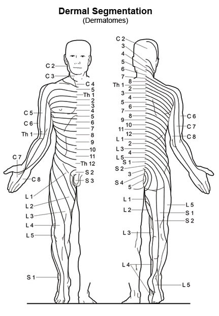 Sensory recordings are accomplished by stimulating nerve endingsmost distally located from the recording electrodes on the scalp. Stimulation sites for nerve roots correspond to a distribution pattern, called dermatomes,* relative to each nerve root (see the diagram below). For example, stimulation of the C6 nerve root is accomplished by stimulating the thumb to send the electrical impulse up through the median (peripheral) nerve to enter the spinal cord at the C6 nerve root located at the C5-C6 level. In order to obtain the most relevant information, stimulate at the location most distal from the area of interest. Peripheral nerves are mixed nerves and are stimulated distally as well. The impulse enters the spinal cord through multiple nerve roots.