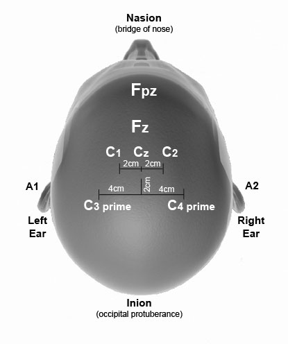
To record somatosensory-evoked potential data during monitoring, subdermal needle electrodes are inserted into the scalp. To acquire motor-evoked potentials, stimulating subdermal or corkscrew needle electrodes must be inserted into the scalp. The International EEG 10/20 Electrode Placement System is referenced below for needle locations. For monitoring purposes, a total of nine electrodes may be used. Six electrodes are used for recording, two electrodes are used for stimulating, and one electrode is used as a ground and may be placed in the scalp (cephalic) or on the shoulder (noncephalic). A grounding pad may be used in lieu of a needle.
The reference points and placement of the electrodes in the average adult are as follows and as depicted in the diagram below:
| Reference Point | Electrode Type | Location | |
|---|---|---|---|
| Fpz | Recording | 10% of the distance from the bridge of the nose (nasion) to the bony knob (occipital protuberance or inion) in the back of the head | |
| Fz | Cephalic ground | Generally between Fpz and Cz | |
| Cz | Recording | 50% of the distance between the nasion and the inion | |
| C1 | Stimulating | 10% of the distance left lateral from Cz or 2 centimeters (cm) left of Cz | |
| C2 | Stimulating | 10% of the distance right lateral from Cz or 2 cm right of Cz | |
| C3 prime | Recording | 20% of the distance left lateral from Cz and 2 cm posterior to Cz or 2 cm posterior and 4 cm left lateral to Cz | |
| C4 prime | Recording | 20% of the distance right lateral from Cz and 2 cm posterior to Cz or 2 cm posterior and 4 cm right lateral to Cz | |
| A1 | Recording | Left ear lobe or mastoid process (bone behind ear) | |
| A2 | Recording | Recording Right ear lobe or mastoid process (bone behind ear) | |
| Additional subdermal needle recording electrodes are inserted in the interspinous spaces between C5-C6 (median nerve) and C7-T1 (ulnar nerve). These locations (not diagramed) represent the root entry zone (median and ulnar nerve origin) to acquire subcortical data. Subcortical data are valuable in evaluating peripheral and central conduction. | |||